Εμφάνιση αναρτήσεων με ετικέτα BIOLOGY OF CANCER. Εμφάνιση όλων των αναρτήσεων

Δευτέρα 4 Ιανουαρίου 2021

RESIDUAL BREAST CANCER BURDEN INDEX

 Residual cancer burden after neoadjuvant chemotherapy can accurately predict disease recurrence and survival across all breast cancer subtypes, according to the findings from a meta-analysis presented at the 2019 San Antonio Breast Cancer Symposium by W. Fraser Symmans, MD, Professor and Director of Research Operations in Pathology at The University of Texas MD Anderson Cancer Center, Houston.1


This is really about organizing the workflow in pathology to standardize how we evaluate response after neoadjuvant treatment.
— W. Fraser Symmans, MD

Tweet this quote

This is not the first study to show residual cancer burden to be an independent factor for prognosis after neoadjuvant chemotherapy. This study, however, goes a step further in estimating the long-term prognosis for each class of residual cancer burden across breast cancer subtypes.

“The most important conclusion is that there is a strong potential to calibrate an individual’s [residual cancer burden] index score to her residual prognostic risk,” Dr. Symmans said. “There is a generally linear relationship between [residual cancer burden] index value and the log of risk.”

Calculating Residual Cancer Burden

“This is really about organizing the workflow in pathology to standardize how we evaluate response after neoadjuvant treatment,” Dr. Symmans said. He explained that pathologists calculate residual cancer burden from multiple factors: primary tumor area, percentage of the tumor area that is invasive cancer, and extent of lymph node involvement. Investigators at The University of Texas MD Anderson Cancer Center put these factors together to develop a residual cancer burden calculator, which computes an index and allocates a classification of pathologic complete response (www.mdanderson.org/breastcancer_RCB). A value of 0 equates to a pathologic complete response. This is then categorized into one of the following three classes: RCB-I (minimal burden), RCB-II (moderate burden), and RCB-III (extensive burden).

KEY POINTS

  • The residual cancer burden index categorizes patients with breast cancer into four groups (RCB 0–IV) based on level of residual disease after neoadjuvant therapy and several other factors as assessed by pathologists.
  • The index has a log-linear relationship with event-free survival at 5 and 10 years.
  • For all subtypes, residual cancer burden tracked consistently with long-term outcome.
  • The residual cancer burden calculator is available to all pathologists through an online calculator (www.mdanderson.org/breastcancer_RCB).

“The basic principle is that we estimate the area that still contains residual disease, map that area to the slides that we’ll be looking at under the microscope, and create an image so that we can reconstruct it and be able to determine what area still contains actual cancer,” Dr. Symmans explained. “Then, we combine that with the fraction of that area that still contains invasive cancer cells, as well as the number of positive lymph nodes and the size of the largest metastasis. This is just organizing what we would otherwise report in pathology, but we’re doing it in a quantitative and standardized manner.”

The online calculator page receives about 16,000 visits per month, “so it’s being used out there,” he noted. The website offers instructional videos, protocols, illustrations, and diagrams as resources for the pathologists who use it.

Pooled Data

The pooled analysis came from the I-SPY Clinical Trials Consortium, involving 12 institutions or clinical trials and encompassing 5,160 patients. The study examined the relationship between the continuous residual cancer burden index and event-free survival, as well as distant relapse–free survival for four breast cancer phenotypes: hormone receptor–negative/HER2-negative (triple-negative), hormone receptor–negative/HER2-positive, hormone receptor–positive/HER2-positive, and hormone receptor–positive/HER2-negative. For each subtype, a multivariate analysis adjusted for patient age, tumor size, nodal status, and grade.

Relationship Between Residual Cancer Burden and Prognosis

The residual cancer burden index was tightly associated with both event-free and distant disease–free survival. This finding was consistent across 12 clinical sites and all cancer subtypes.

“The most interesting result, in my opinion, is that you see a log-linear relationship for the [residual cancer burden] index score and survival,” Dr. Symmans said. “The implication is that you can take an individual patient’s score representing how much residual cancer she has and calibrate that to an accurate estimate of her risk over time.”

A pathologic complete response (RCB-0) was most likely to be achieved by hormone receptor–negative/HER2-positive patients (69%) and least likely by the hormone receptor–positive/HER2-negative group (11%); the triple-negative group (43%) and hormone receptor–positive/HER2-positive group (38%) fell in between.

The residual cancer burden index values were associated with 5-year and 10-year event-free survival, as shown in Table 1.

Elaborating on what emerged from the four subgroups, Dr. Symmans said that in the hormone receptor–negative/HER2-positive group, the 69% rate of pathologic complete response was “striking” and “illustrates how effective these current [HER2-targeted] treatments are.” The 20% of patients who were classified as RCB-II and RCB-III had significantly worse survival than the others. “It’s a small quintile of patients that’s still at fairly substantial risk.”
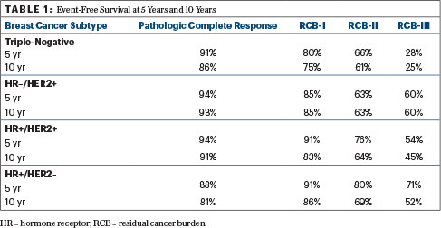
He added, “In the hormone receptor–positive/HER2-negative breast cancer group, where there is still a bit of confusion about whether or not chemotherapy can help patients, we see that the extent of residual disease is strongly prognostic.”

In the subgroup with hormone receptor–positive/HER2-negative disease, he continued, “you can see that there’s clearly an effect on prognosis from the chemotherapy. However, the most prognostic aspect of the distribution of response is where there is the most residual disease—the RCB-III and RCB-II groups. Their long-term risk is still continuing beyond 10 years.”

The residual cancer burden index remained independently prognostic in multivariate models adjusting for patient age, grade, and clinical T and N stage at diagnosis. Its value was 1.93 in the triple-negative group, 2.04 in the hormone receptor–negative/HER2-positive group, 1.67 in the hormone receptor–positive/HER2-positive group, and 1.52 in the hormone receptor–positive/HER2-negative group. T4 tumors (and in the triple-negative group, also T3) remained independent predictors of prognosis as well.

Message to the Pathology Community

“Looking ahead, if we can standardize the reporting of residual cancer burden, that will only improve its usefulness in determining long-term prognosis,” Dr. Symmans emphasized. “This study is important in showing the generalizability of residual cancer burden, and it’s of sufficient size to generate the evidence to convince the pathology community to change the way we do things.”

Looking ahead, if we can standardize the reporting of residual cancer burden, that will only improve its usefulness in determining long-term prognosis.
— W. Fraser Symmans, MD

Tweet this quote

Dr. Symmans noted that synoptic reporting is a requirement in oncology, but there is only one synoptic report for invasive cancer—one that is designed for a surgery-first examination. What’s lacking is a neoadjuvant synoptic report that specifically addresses the needs in interpreting a post-neoadjuvant response, he said. “The neoadjuvant model is increasingly related to precision medicine. We are standing at that transition point.” 

DISCLOSURE: Dr. Symmans holds a patent for the method of calculating residual cancer burden. The residual cancer burden calculator and educational materials are freely and publicly available online. He is also cofounder, owns shares in, and is an unpaid scientific advisor for Delphi Diagnostics.

REFERENCE

Κυριακή 20 Δεκεμβρίου 2020

CHEMOTHERAPY BENEFICIAL IN ELDERLY WITH TNBC

 In a National Cancer Database analysis reported in The Lancet Oncology, Crozier et al found that receipt of neoadjuvant or adjuvant chemotherapy was associated with improved overall survival among women aged 70 or older undergoing surgery for stage I to III triple-negative breast cancer.

Study Details

The analysis included data from the National Cancer Database from women aged 70 or older with surgically treated stage I to III invasive triple-negative breast cancer diagnosed between 2004 and 2014. Patients with T1a N0 M0 disease and those with incomplete data on estrogen receptor, progesterone receptor, or HER2 status were excluded from the analysis.

Patients were categorized into three groups: those who received chemotherapy (chemotherapy group), those who were recommended chemotherapy but did not receive it (recommended/not received group), and those for whom chemotherapy was not recommended and not received (not recommended/not received group).

Overall Survival in Total Cohort

KEY POINTS

  • Receipt of chemotherapy in addition to local therapy was associated with significantly improved overall survival.
  • Benefit was observed in node-negative and node-positive disease and in patients with comorbidities.

A total of 16,602 women were included in the analysis. Of these, 7,485 (46.6%) received chemotherapy; chemotherapy was recommended for but not administered to 2,659 (16.6%); chemotherapy was not recommended for or received by 5,732 (35.7%); and chemotherapy status was not available for 186 (1.2%). Of the patients who received chemotherapy, 5,924 (79.1%) received adjuvant chemotherapy alone; 1,337 (17.9%) received neoadjuvant chemotherapy alone; and 196 (2.6%) received both.

Median follow-up was 38.3 months (interquartile range = 20.7–46.1 months, range = 0–138.0 months).  At data cutoff at the end of January 2016, 3,300 patients (20.5%) had died.

The 5-year overall survival estimate for the entire cohort was 62.3% (95% confidence interval [CI] = 59.7%–64.4%). The 5-year estimates were 68.5% (95% CI = 66.4%–70.6%) in the chemotherapy group, 61.1% (95% CI = 59.0%–63.2%) in the recommended/not received group, and 53.7% (95% CI = 51.8%–55.8%) in the not recommended/not received group (overall < .0001).

On univariate analysis, hazard ratios were 0.80 (95% CI = 0.73–0.88, P < .0001) for the recommended/not received group and 0.58 (95% CI = 0.54–0.62, P < .0001) for the chemotherapy group vs the not recommended/not received group. There was no apparent correlation of year of diagnosis with overall survival (P = .48).

On multivariate analysis, hazard ratios remained significant at 0.79 (95% CI = 0.71–0.88, P < .0001) and 0.56 (95% CI = 0.51–0.61, P < .0001), respectively. Patients who received radiotherapy (48.7% of entire cohort) also had improved overall survival compared with those who did not receive radiotherapy (50.0% of cohort; HR = 0.62, 95% CI = 0.50–0.79, < .0001).

Propensity Score–Matching Analysis

A propensity score–matching analysis was performed that included 1,884 patients who received chemotherapy and 1,884 patients in the recommended/not received group, with matching based on age, comorbidity score, tumor grade and size, nodal status, and receipt vs no receipt of radiotherapy.

Estimated 5-year overall survival was 66.8% (95% CI = 65.7%–67.9%) in the chemotherapy group vs 61.8% (95% CI = 60.8%–62.9%) in the recommended/not received group (HR = 0.85, 95% CI = 0.74–0.96, = .012). The difference remained significant on multivariate analysis (HR = 0.69, 95% CI = 0.60–0.80, < .0001). There was no clear indication that the benefit of chemotherapy was limited to any age subgroup.

In subset analyses in the propensity-matched cohort, estimated 5-year overall survival among women with node-negative disease (74% vs 66% of cohort, all with tumor size > 5 mm) was 74.4% (95% CI = 72.6%–76.2%) in the chemotherapy group vs 70.7% (95% CI = 68.9%–72.4%) in the recommended/not received group (HR = 0.80, 95% CI = 0.66–0.97, = .007). Estimated 5-year overall survival among women with node-positive disease was 42.4% (95% CI = 39.9%–44.9%) vs 35.1% (95% CI = 32.6%–37.6%; HR = 0.76, 95% CI = 0.64–0.91, = .006). Among women with a documented Charlson-Deyo comorbidity score greater than 0 (25% vs 26% of cohort), the hazard ratio was 0.74 (95% CI = 0.59–0.94, = .013).

On multivariate analysis in the propensity-matched cohort, additional factors significantly associated with overall survival included:

  • Age as a continuous variable (HR = 1.03, P = .0010)
  • Comorbidity score of 1 (HR = 1.54, P < .001) and 2 (HR =1.38, P = .011) vs 0
  • Tumor size > 50 mm vs ≤ 5 mm (HR = 4.38, P = .042)
  • pN1, pN2, and pN3 disease (HRs = 1.92, 3.26, and 5.59, all P < .0001) vs pN0 disease
  • Receipt vs no receipt of radiotherapy (HR = 0.62, P < .0001).

The investigators concluded: “These data support consideration of chemotherapy in the treatment of women aged 70 years or older with triple-negative breast cancer.”

Christopher M. Pezzi, MD, of the Division of Surgery, Baptist MD Anderson Cancer Center, Jacksonville, is the corresponding author for The Lancet Oncology article

LESS WOMEN WITH BREAST CANCER TO RECEIVE AADJUVANT CHEMO-RxPONDER TRIAL

 More women with early-stage breast cancer may safely forgo chemotherapy, suggests an interim analysis that had a median follow-up of 5 years of the large-scale phase 3 RxPONDER trial, presented this week at the San Antonio Breast Cancer Symposium (SABCS) 2020.

The investigators reported that adding chemotherapy to endocrine therapy did not improve outcomes for postmenopausal women with low-risk, node-positive, hormone receptor–positive (HR+), human epidermal growth factor receptor 2–negative (HER2-) breast cancer in comparison to endocrine therapy alone.

These results are akin to those from the TAILORx trial. The results of that trial were first presented in 2018 and have changed practice for women with early-stage disease who have no lymph node involvement.

Clinicians celebrated the new results for women with lymph node–positive disease.

"RxPonder: practice changing!!!" tweeted meeting attendee Sarah Sammons, MD, Duke Cancer Center, Durham, North Carolina.

"Data from RxPonder are the most clinically important this year at @SABCSSanAntonio," tweeted Hal Burstein, MD, Dana Farber Cancer Institute, Boston, Massachusetts, who was not involved in the study.

"This will save tens of thousands of women the time, expense, and potentially harmful side effects that can be associated with chemotherapy infusions," asserted study lead author Kevin Kalinsky, MD, Winship Cancer Institute of Emory University, Atlanta, Georgia, during a meeting press conference.

But the trial, run by the SWOG Cancer Research Network, was not without controversy.

That's because the trial also included premenopausal women whose disease characteristics were the same and who were found to have benefited from chemotherapy.

It was not clear whether the benefit was from chemotherapy's cytotoxicity or its endocrine effects/ovarian suppression (which limits the production of estrogen, a breast cell stimulant) in these young women. But multiple experts asserted that the effect was very likely from ovarian suppression. "There are less toxic ways than chemo to suppress ovarian function," tweeted Tatiana Prowell, MD, Johns Hopkins University, Baltimore, Maryland, who is not a study investigator.

Some experts strongly doubted the findings in premenopausal women.

"I hate to come away with the message that all [low-risk, node-positive] premenopausal patients should get chemotherapy," summarized C. Kent Osborne, MD, Baylor College of Medicine, Houston, Texas, who is co-director of SABCS and was not involved in the study.

RxPONDER will follow patients for 15 years, so additional data and insights will follow, observed SWOG in a press statement.

Women Had Limited Positive Nodes

RxPONDER, or SWOG S1007, involved more than 5000 women who had HR+, HER2- breast cancer with involvement of one to three lymph nodes. The patients' recurrence score was ≤25 on a 21-tumor gene expression assay (Oncotype Dx), which is characterized as low risk.

Approximately 20% of US women with nonmetastatic HR+, HER2- breast cancer present with involvement of one to three lymph nodes, added Kalinsky.

Study participants were randomly assigned to receive either standard chemotherapy plus endocrine therapy or endocrine therapy alone. Follow-up was for a median of 5 years before the current preplanned analysis.

Over a median follow-up of 5.1 years, there were 447 observed invasive disease-free survival (IDFS) events, the primary endpoint, which is 54% of the expected number at final analysis.

Across the whole cohort, adding chemotherapy to endocrine therapy was associated with a significant improvement in IDFS, with a 5-year rate of 92.4% vs 91.0% for endocrine therapy (= .026).

Among the postmenopausal women, no such improvement was seen. The 5-year IDFS rate was 91.6% with chemotherapy plus endocrine therapy and 91.9% with endocrine therapy alone (= .82).

Among premenopausal women, there was improvement in IDFS. The 5-year rate was 94.2% with chemotherapy plus endocrine therapy, vs 89.0% for endocrine therapy alone (= .0004).

These differences were reflected in the results for overall survival. For postmenopausal women, there was a nonsignificant difference in 5-year overall survival rates (96.2% vs 96.1%).

On the other hand, for premenopausal women, there was a significant difference in 5-year overall survival rates (98.6% vs 97.3%; = .032).

Stratifying patients by recurrence score 0–13 vs 14–25 and by involvement of one vs two to three nodes did not have a major impact on the results, said lead author Kalinsky, who also noted that future analyses will include quality of life and other outcomes.

More About Endocrine Therapy in RxPONDER

Meeting co-director Osborne said that premenopausal women in RxPONDER were "nearly always" prescribed tamoxifen.

However, he observed that the current standard approach to treatment in this age group would be ovarian suppression plus either an aromatase inhibitor or tamoxifen, "both of which have been shown to be superior to tamoxifen alone in this subgroup.

"Since the adjuvant chemotherapy causes ovarian suppression in many premenopausal patients," he said, "these patients then in fact received ovarian suppression plus tamoxifen," rather than tamoxifen alone for the group that did not receive chemotherapy.

Osborne asked a question that came up again and again during the post-presentation discussion: "Is the difference in outcome in this subset due to the endocrine effects of chemotherapy?

"Unfortunately, we may never know the answer to this question," he added.

Kalinsky replied that whether the difference in benefit of chemotherapy in premenopausal women "was a direct benefit, meaning that there's something about the biology difference" between tumors in premenopausal vs postmenopausal women, "or whether this was an indirect effect, meaning impacting rates of amenorrhea...is not specifically how this study was designed."

However, an exploratory landmark analysis at 6 months suggested that the use of ovarian suppression with endocrine therapy did not have an effect on outcomes.

Osborne said he is nevertheless "still skeptical that chemotherapy works differently in premenopausal women.

"Until we show that it's not an endocrine effect...I just can't imagine why that group of patients, even the ones with very low Oncotype [score], would have a different response to chemotherapy."

He added: "If I can think of a rationale...I would believe it, but right now, I'm a little bit skeptical."

Virginia Kaklamani, MD, of the UT Health San Antonio Cancer Center, San Antonio, Texas, who is a meeting co-director, said that she wanted to "second that.

"I honestly think that this is an OFS [ovarian function suppression effect] that we are seeing. We have several clinical trials that have been done looking at ovarian function suppression vs not...showing that [it] can help as much as chemotherapy."

Kaklamani continued: "Unfortunately, the arms to those trials were not perfect for now, and this is going to be an unanswered question until we have a large trial comparing OFS to chemotherapy."

The study was sponsored by the National Cancer Institute and in part by the Susan G. Komen for the Cure Research Program, the Hope Foundation for Cancer Research, the Breast Cancer Research Foundation, and Exact Sciences. Kalinsky, Osborne, and Kaklamani report financial ties to multiple pharmaceutical companies.

San Antonio Breast Cancer Symposium (SABCS) 2020: Abstract GS3-00. Presented December 10, 2020.

For more from Medscape Oncology, join us on Twitter and Facebook.

CTCs TO GUIDE BREAST CANCER THERAPY-NOT YET

 Trial results suggest circulating tumor cell (CTC) counts may be a reliable biomarker for guiding the choice of first-line treatment in patients with hormone receptor–positive, HER2-negative metastatic breast cancer, investigators wrote in JAMA Oncology.

However, authors of a related editorial suggested CTC counts are not adequate for guiding treatment choice in this population.

In a phase 3 trial, investigators compared the use of CTC counts and the use of clinical factors to guide the decision between chemotherapy and endocrine therapy. Results showed similar progression-free survival (PFS) and overall survival (OS) with both methods but more chemotherapy use with the CTC method.

"The results of this trial demonstrate the reliability and clinical utility of CTC count to guide the choice between single-agent endocrine therapy and chemotherapy as first-line treatment," but "at the cost of a higher proportion of patients treated with chemotherapy," study author François-Clement Bidard, MD, PhD, of Institut Curie in Saint-Cloud, France, and colleagues wrote.

The investigators explained that endocrine therapy is the preferred first-line treatment option in this patient population, but chemotherapy is used when women are in visceral crisis, with rapidly progressive, symptomatic disease. The decision usually rests on clinical factors, such as tumor subtype and performance status, but there's interphysician variability.

The team hoped to find a "more reliable, standardized, and reproducible" biomarker to help remove some of the uncertainty from the situation. They tested CTC count, a well-established prognostic indicator of PFS and OS, as a candidate.

Study Results

The trial included 755 patients with hormone receptor–positive, HER2-negative breast cancer in the per-protocol population. The patients' median age was 63 years (range, 30-88 years).

Among the 377 patients randomized to the CTC arm, those with counts at or above 5 CTCs per 7.5 mL received chemotherapy, while those with a lower count received endocrine therapy.

The 378 patients in the standard-care group received endocrine therapy or chemotherapy based on provider choice guided by clinical factors.

Chemotherapy was given to 37% of patients in the CTC arm and 27% of those in the standard arm.

The median PFS was 15.5 months in the CTC arm and 13.9 months in the standard arm, which meant the primary endpoint of noninferiority was met (hazard ratio, 0.94; 90% confidence interval, 0.81-1.09).

Age older than 60 years was the only baseline characteristic associated with better PFS with CTC-driven decision-making. This may be because of the greater "use of endocrine therapy as the clinically favored treatment, whatever the other clinicopathologic characteristics," in older subjects, the investigators wrote.

As with PFS, the median OS was similar between the study arms – 47.3 months in the CTC arm and 42.8 months in the standard arm (HR, 0.91; 95% CI, 0.71-1.16).

"Not Good Enough"

The investigators behind this study had "a worthy goal," according to authors of a related editorial.

Without "predictive biomarkers, we are left with our clinical knowledge, experience, and intuition. Patients are left with uncertainty, doubt, and fear," Tarah Ballinger, MD,, of Indiana University, Indianapolis, and colleagues wrote in the editorial.

However, the editorialists had concerns about the findings. For one thing, the investigators hypothesized that relying on CTC would lead to a deescalation from chemotherapy to endocrine therapy, but use of chemotherapy was actually 10% higher in the CTC arm.

"Adding to or replacing the parameters we use to make a clinical decision should help us improve the lives of patients. ... We should demand an improvement in outcomes before accepting a strategy that exposes more patients to more toxic therapy. Not worse simply is not good enough," the editorialists wrote.

In addition, the trial was completed before CDK4/6 inhibitors became a standard add-on with endocrine therapy for hormone receptor–positive, HER2-negative patients.

"The overall response rate to CDK4/6 inhibitor therapy is higher than with traditional chemotherapy, and several randomized trials have failed to show a survival benefit of upfront chemotherapy compared with CDK4/6 inhibitor use. ... Thus, it is even less likely that we can assume that baseline high CTC count corresponds to a need for chemotherapy in a modern treatment landscape that offers more patients more benefit from hormone therapy," Ballinger and colleagues wrote.

The editorialists concluded that CTC count "alone at baseline primarily reflects disease bulk, much like anatomic staging, rather than disease biology. As treatments become more rooted in our knowledge of breast cancer biology, decisions based on disease bulk are decidedly out of place."

Perhaps a better use, they suggested, is for treatment personalization. For instance, patients with persistently elevated CTCs despite standard approaches could consider trials of novel targeted therapies, or CTCs could be sequenced to identify actionable molecular targets, achieving a "clinical utility that merely counting CTCs lacks," the editorialists wrote.

This study was funded by the Institut Curie, the French National Cancer Institute, and Menarini Silicon Biosystems, the maker of the CTC assay used in the trial. The investigators disclosed relationships with Menarini and many other companies. Ballinger receives honoraria from Medscape, which is owned by the same company as this news organization.

SOURCE: Bidard FC et al. JAMA Oncol. 2020 Nov 5. doi: 10.1001/jamaoncol.2020.5660.

Δευτέρα 7 Δεκεμβρίου 2020

EXCEPTIONAL RESPONDERS TO CANCER TREATMENT

A small percentage of patients with cancer show exceptional responses to treatment and survive significantly longer than patients with clinically comparable tumors, despite having advanced disease.

An ongoing research project is studying why some patients have exceptional responses. The researchers have found particular molecular features in the tumors of about a quarter of these patients. In some cases, there are multiple rare genetic changes in the tumor genome. In other cases, the tumors are infiltrated with certain types of immune cells.

The findings were published online November 19 in Cancer Cell. They come from a genomic analysis of tumor biopsy specimens from 111 patients who were identified by the National Cancer Institute's (NCI's) Exceptional Responders Initiative, a national project launched in 2014.

An exceptional responder is defined as an individual who achieves a partial or complete response to a treatment that would be effective in fewer than 10% of similar patients. For exceptional response, the duration of response is at least three times longer than the usual median response time.

In this study of 111 such patients, about one quarter (24%, n = 26 patients) were found to have tumors in which there were molecular features that could potentially explain exceptional responses to treatment.

"We won't be able to identify, in every patient, which particular drugs will be beneficial," said Louis Staudt, MD, PhD, director of the NCI's Center for Cancer Genomics, who co-led the study. "We are nowhere near that. But what it does say is that we have identified particular mutations, some of which we knew about in some types of cancer but can also occur less commonly in other cancer types."

Staudt noted that these mutations can "illuminate" the path that the cancer will take — and potentially can be used to predict whether the cancer will be aggressive and will require treatment or could be managed with surveillance. This is why this research can be useful in the short term, he said.

"In the longer term, this is the kind of research that inspires future work," he told Medscape Medical News. "That would encompass clinical trials involving drugs that target some of the pathways we found to be genetically inactivated in some of these responders."

These results support the use of genetic testing in routine clinical care, he said.

Earlier this year, the NCI team published the results of a pilot study that affirmed the feasibility of this approach. Of the more than 100 cases that were analyzed, six were identified as involving potentially clinically actionable germline mutations.

"Curiosity Drove the Research"

"We had these wonderful and gratifying experiences with our patients, so we were immediately curious how that happened, so it was pretty much that curiosity that drove a lot of this work," said Staudt.

In the current study, Staudt and colleagues used multiple genomic methodologies to detect mutations, copy number changes, aberrant methylation, outlier gene expression, and the cellular makeup of the tumor microenvironment.

The hypothesized mechanisms for exceptional responses were broadly divided into the following four categories: DNA damage response (n = 15), intracellular signaling pathway (n = 9), prognostic genetics (n = 9), and immunologic engagement (n = 16). For many patients, two or more of these mechanisms were involved.

The authors note that the "predominance of plausible DNA damage response mechanisms parallels the frequent use of cytotoxic chemotherapy in routine cancer treatment reflected in this cohort."

Twenty-six patients were identified as exceptional responders. Among these patients, a variety of cancer types was represented: brain (8); gastrointestinal tract (6); breast (4); cholangiocarcinoma (2); lung (2); pancreas, endometrium, ovarian, and bladder (1 each). Many of these patients (65%, n = 17) were treated with chemotherapy that included DNA-damaging agents. For more than half (54%, n = 14), targeted therapies were used, and some patients received both.

The authors highlight several patients as examples of exceptional responders:

  • One patient with glioblastoma multiforme (GBM) was treated sequentially with surgery, localized carmustine, and radiotherapy. When the cancer recurred, temozolomide was administered. This induced a complete response that has lasted for more than a decade.

  • A patient with metastatic colon adenocarcinoma has had an ongoing and nearly complete response that has lasted 45 months (last follow-up) after receiving temozolomide in combination with the investigational drug TRC102 (methoxyamine, under development by Tracon) in a phase 1 clinical trial. TRC102 is an inhibitor of the DNA base excision repair pathway, which is a pathway that causes resistance to alkylating and antimetabolite chemotherapeutics.

  • A patient with metastatic, estrogen receptor–positive breast cancer received trastuzumab because of a high-level ERBB2 amplification, together with anastrozole (Arimidex). This resulted in an ongoing 2.4-year partial response.

    Although the patient was clinically HER2 positive, her tumor had exceedingly low expression of ERBB2 mRNA. Molecular profiling had classified the tumor as of the basal-like subtype rather than the HER2-enriched subtype. This meant that it was unlikely that trastuzumab contributed to the exceptional response, the authors note. Because the patient was estrogen-receptor positive, she received anastrozole, an inhibitor of aromatase (CYP19A), which converts testosterone into estradiol.

  • A patient with a gastrointestinal stromal tumor with a deletion of KIT exon 11 experienced relapsed after an initial response to imatinib, which targets KIT, but then achieved a complete response with sunitinib (Sutent). Gene expression profiling revealed high expression not only of KIT but also of genes encoding several tyrosine kinases that are targeted by sunitinib (KDR, FLT1, and FLT3). This may have accounted for the patient's response.

Favorable Genomic Characteristics

The authors defined a "prognostic genetics" category of tumors, characterized by genetic lesions that are now known to be associated with a favorable prognosis but that were not addressed through routine care that these patients received when they were first diagnosed. Although the patients experienced relapse after first-line treatment, their exceptional survival after salvage therapy could be linked to favorable genomic characteristics.

For example, several the patients with high-grade GBMs and astrocytomas had genetic lesions that are generally more common in low-grade glioma and that have been associated with an indolent clinical course following standard therapy.

The authors also assessed immune response. Examining immune cell infiltration in responder tumors in comparison with control cases, they found that signatures of B cells and activated (CD56dim) natural killer cells were higher in exceptional-responder tumors. In one patient with metastatic urothelial cancer who experienced disease progression after chemotherapy, radiotherapy, and surgery, treatment with nivolumab (Opdivo) produced a complete response that lasted 7 months. Such an outcome occurs in only about 3% of bladder cancer patients. The tumor expressed high mRNA levels of PDCD1, which encodes the nivolumab target PD-1, and CD274, which encodes the PD-1 ligand PD-L1. There was also a high level of amplification of IFNG, which encodes interferon-gamma, a cytokine that has been linked to favorable response to immune checkpoint blockade.

Moving to Precision Medicine

"It is very valuable to be tested up front and again when the disease progresses, because there may have been some genetic changes, and this may change the treatment," said co–lead author S. Percy Ivy, MD, of the NCI's Division of Cancer Treatment and Diagnosis.

"The goal of this study was to understand what was unique about these patients and their genetic makeup that led them to be classified as exceptional responders, and hopefully we will be able to tease that out," she added.

"As researchers, we have a lot to learn from these patients, and they have a lot to teach us," she added. "In the future, they will help us as we move closer to the goal of delivering precision oncology to all of our patients. We're not there yet, but every time we study more deeply and learn more, we are able to provide better care."

To encourage participation in this effort by investigators around the world, the NCI team and their colleagues have made their molecular profiling results and clinical information publicly available in the NCI Genomic Data Commons.

The study was supported by the NCI's Intramural Research Program, the National Institutes of Health, the Center for Cancer Research, and the NCI's Center for Cancer Genomics. Staudt and Ivy have disclosed no relevant financial relationships.

Cancer Cell. Published online November 19, 2020. Abstract

For more from Medscape Oncology, join us on Twitter and Facebook.

Medscape Medical News © 2020


LIQUID BIOPSIES NOT ALWAYS THE ANSWER

 In men with prostate cancer, cell-free DNA (cfDNA) testing may be confounded by clonal hematopoiesis (CHIP) gene variants, leading some patients to be treated inappropriately with poly(ADP) ribose polymerase (PARP) inhibitors.

"Our study highlights an important limitation of most current cfDNA or 'liquid biopsy' testing for men with prostate cancer," Dr. Colin C. Pritchard of the University of Washington, in Seattle, told Reuters Health by email. "We found that blood-cell mutations were commonly detected in blood plasma, and that these mutations could be misinterpreted as coming from prostate cancer. The results have important implications for guiding therapy with a newer class of drugs called PARP inhibitors."

"Almost half the time that mutations related to (U.S. Food and Drug Administration)-approved indications for PARP inhibitor eligibility were detected in cfDNA, they were derived from blood-cell clonal hematopoiesis, and not related to prostate cancer," he said. "These interfering clonal hematopoiesis mutations were particularly common in older age groups."

Dr. Pritchard and colleagues examined 69 men with advanced prostate cancer who underwent cfDNA variant testing. To determine the source of variants, they also tested paired whole blood control samples.

Twenty patients showed pathogenic variants in relevant DNA repair gene variants, the researchers report in JAMA Oncology. CHIP variants at 2% or more variant fraction in cfDNA were seen in 13. Of these men, seven had variants in DNA-repair genes used to determine PARP inhibitor candidacy and would have been misdiagnosed and incorrectly deemed eligible for PARP-inhibitor therapy.

In fact, unknown to the researchers at the time, one of this latter group had already had a recommendation for PARP-inhibitor therapy from a commercial laboratory which used only plasma testing.

"Fortunately," continued Dr. Pritchard, "there is a simple solution to improve the performance of cfDNA tests. We found that by incorporating a paired whole-blood control sample into the test we were easily able to distinguish prostate-cancer mutations in plasma cfDNA from blood-cell clonal hematopoiesis. We have been using this paired testing approach for cfDNA testing . . . but most of the currently available commercial cfDNA assays test only plasma, and cannot readily distinguish cancer mutations from blood cell interference."

"For this reason," he concluded, "we are concerned that men with the most advanced forms of prostate cancer may be incorrectly receiving PARP inhibitor therapy based on false positive cfDNA testing results."

Dr. Joshi J. Alumkal of the University of Michigan, in Ann Arbor, who co-wrote an accompanying editorial, told Reuters Health by email that the results "provide a cautionary note on using cell-free DNA sequencing to identify patients with mutations in homologous recombination DNA-repair genes - gene mutations that are routinely used for PARP inhibitor treatment selection."

He added, "Understanding whether these mutations are due to tumor cells versus clonal hematopoiesis is critical to make the most well-informed decisions about patient management."

SOURCE: https://bit.ly/3n2pQCm and https://bit.ly/35eWOtp JAMA Oncology, online November 5, 2020.

Reuters Health Information © 2020

DIAGNOSTIC TESTS AND RISK OF TESTICULAR CANCER

Over the last 40 years, there has been a doubling in the incidence of testicular cancer. Over the same period, there has been a 20-fold increase in the use of diagnostic radiation, and the doses of radiation used have increased sevenfold. Perhaps the two are related?

The suggestion comes from researchers reporting a new case-control study in which they show that repeated diagnostic radiation below the waist was associated with an increase the risk of testicular germ cell tumor (TGCT).

The study involved 315 men with TGCT and 931 controls from hospital and population-based settings, and analyzed medical records and individuals'  recall of diagnostic radiation such as CT scans, x-rays, and barium enema.

Compared with men who did not undergo any diagnostic radiation, the risk of testicular cancer was nearly twofold higher among men who had undergone three or more diagnostic X-rays and CT scans, and more than fourfold higher among men who had undergone three or more lower gastrointestinal (GI) series or barium enema.

The study was published online November 11 in PLoS ONE.

Testicular cancer, the most common cancer in white men aged 15-44, has steadily increased in incidence in the United States over the past three or four decades: the rate has jumped from 3 out 100,000 men in 1975 to 6 out of 100,000 men today, say senior author Katherine Nathanson, MD, of the University of Pennsylvania in Philadelphia, and colleagues.

"Our data suggests that the increased use of diagnostic radiation below the waist in men over that same time may contribute to the increase in incidence," Nathanson said in a press statement.

Reducing diagnostic radiation doses to the testes "should be prioritized," say the investigators, if these results are validated by additional research.

It's "undoubtedly prudent" to reduce those doses "as much as possible," said David Brenner, PhD, director, Center for Radiological Research, Columbia University Medical Center, New York City, who was asked for comment.

I do find the results quite surprising. Dr David Brenner

However, Brenner also had reservations about the new study. "I do find the results quite surprising," given what is known historically about testicular cancer and radiation exposure, he said.

Higher radiation doses (than in the current study) in two other settings (among post-atomic bomb survivors and in the treatment of ankylosing spondylitis) are not associated with an increased risk of testicular cancer, he told Medscape Medical News in an email.

"Underlying conditions for which the individual received radiation imaging," may be one potential explanation for the increased testicular cancer risk seen in this study, he added.

Nathanson responded to Brenner's critique by focusing on atomic bomb survivors and pointing out that the baseline rate of testicular cancer in Asia, including Japan, is "very, very low" and therefore that population, including bomb survivors, is not a highly applicable comparison.

Take-Home Message: Testicular Shielding

Nathanson noted that diagnostic imaging below the waist is most commonly administered as a result of GI tract complaints such as constipation and abdominal pain.

For clinicians, the study's take-home message supports testicular shielding, she told Medscape Medical News.

Testicular shielding during diagnostic imaging can be protective, but audits show that correct use and positioning occurs in just 25% of pediatric diagnostic scans, the authors comment.

Recent epidemiologic studies support a tie between diagnostic radiation and other cancers, the authors also point out.

For example, women undergoing frequent x-rays for scoliosis may have an increased risk of breast cancer, and likewise for individuals undergoing repeat routine dental x-rays and thyroid cancer. Additionally, a National Health Service study among 175,000 children revealed an excess risk of subsequent brain tumors and leukemia tied to CT scans, say Nathanson and coauthors.

Total-dose and number of exposures were associated with an increasing cancer risk in each of the aforementioned studies.

Nathanson pointed out that Penn researchers have collected testicular cancer data since 2001 for the sake of research on related, inherited risk. The new study is an outgrowth of those efforts and is a rare effort in this subject area, in part, because testicular cancer is a very treatable rare cancer, with a 95% survival rate at 5 years.

A larger prospective study of the association between diagnostic radiation and testicular cancer would be better for determining causality, but is impractical given the needed time and associated costs. "A case-control design is a methodology that is ideally suited for studies investigating rare disease outcomes such as TGCT," the authors comment.

The study was supported with grants from the National Cancer Institute and the National Institutes of Health. The study authors and Brenner have disclosed no relevant financial relationships.

PLoS One. Published online November 11, 2020. Full text

Nick Mulcahy is an award-winning senior journalist for Medscape. He previously freelanced for HealthDay, MedPageToday and has had bylines on WashingtonPost.com, MSNBC, and Yahoo. Email: nmulcahy@medscape.net and on Twitter: @MulcahyNick


Κυριακή 15 Νοεμβρίου 2020

TEST ALL CANCER PATIENTS FOR DRIVER MUTATIONS

About 1 in 8 patients with cancer have inherited genetic mutations that may have contributed to the development of their cancers, but nearly half of these mutations would have been missed using current clinical guidelines.

These findings come from the largest study of its kind so far, conducted in nearly 3000 patients with a wide range of cancer stages and types, including breast, colorectal, lung, ovarian, pancreatic, bladder, prostate, and endometrial cancers.

"This study tells us that the clinical practice guidelines are not very sensitive for identifying who does or doesn't have a genetic mutation that is predisposing them to cancer," commented first author Niloy Jewell Samadder, MD, director of the high-risk cancer clinic at the Mayo Clinic in Arizona, Phoenix, Arizona.

Finding a genetic mutation can alter clinical management of the cancer.

"This really does open up treatment and management options that might not have been accessible to these patients," Samadder emphasized.

The results were published online on October 30 in JAMA Oncology and were presented simultaneously at the Society of Human Genetics. Samadder discusses details of the study in a video posted on YouTube.

A clinician not involved in the study said the new results should lead to changes in practice.

"For cancer patients, I think the debate is over. We should test everybody," Peter Beitsch, MD, surgical oncologist at the Dallas Surgical Group, told Medscape Medical News.

The Mayo Clinic is changing its daily practice at all four of its cancer centers. The changes will begin in the first quarter of 2021 at its Arizona campus.

"Every cancer patient who comes to Mayo Clinic will be offered genomic evaluation that includes genetic testing to identify if they have an underlying genetic mutation that predisposes to their cancer and [helps physicians decide] how to incorporate that knowledge into designing the best surgical and treatment options for that patient and their family," Samadder said.

Study Details

The study included 2984 patients with cancer who were receiving care for a variety of solid tumor cancers at Mayo Clinic cancer centers in Arizona, Florida, Minnesota, and a community cancer center in Wisconsin.

Patients were tested for about 84 genes using next-generation sequencing provided by Invitae.

Among participants, 13.3% (n = 397) tested positive for pathogenic mutations. Of these, about 70% (282 of 397 patients) carried moderate- and high-penetrance genes that increased their risk for cancer. For almost 28.2% (n = 42) of patients with high-penetrance mutations, changes were made in treatment as a result of genetic testing. These included changes in surgical management, immunotherapy, chemotherapy, or enrollment in a clinical trial for which they may otherwise have not been eligible.

Researchers also compared their universal testing approach to targeted testing recommended in guidelines from the National Comprehensive Cancer Network, the National Society of Genetic Counselors, and the American College of Medical Genetics.

They identified pathogenic mutations in 192 patients whose mutations would have been missed using guideline-recommended criteria, such as tumor pathology or family history. This represents 6.4% of all participants in the study (192 of 2984 patients) and 48.4% of patients who tested positive for pathogenic mutations (397 of 2984 patients).

"Genetic testing is underutilized in cancer care, both for patients and for their families, often due to outdated guidelines that restrict testing to a narrow group of high-risk patients. All cancer patients should have access to complete genetic information that can guide their care and inform their families' health," coauthor Robert Nussbaum, MD, chief medical officer of Invitae, said in a statement.

Some clinicians have been pushing for genetic testing of all patients with cancer, including Beitsch, who was lead author of a similar study in breast cancer patients published last year in the Journal of Oncology. That article made waves when the authors concluded that all breast cancer patients should have expanded panel genetic testing.

This new Mayo Clinic study extends the findings in breast cancer to "all cancer patients, not just breast cancer patients," Beitsch told Medscape Medical News.

Long-Running Debate

The new findings and opinions add to a long-running debate in oncology over the role of genetic testing and screening for pathogenic mutations.

Part of the debate about genetic testing has hinged on the question of costs, says Beitsch. When genetic testing first became available, it was conducted by hand, and costs were often prohibitive. Since then, genetic testing has been automated using next-generation sequencing, and the cost has decreased considerably.

"The Invitae cash price for an 80-plus gene panel is $250. That's [the cost of] a mani-pedi in Dallas. I don't discount that it's a lot of money for a lot of people. Yes, it's expensive, but it's a lot less expensive than it used to be," Beitsch said.

Another issue is that doctors are not entirely sure how to manage variants of uncertain significance (VUSs) when they are found. In the Mayo Clinic study, about half (47.4%; n = 1415) of participants had VUSs. The authors note that these results are consistent with past studies.

Beitsch says VUSs are a matter of education. To date, only about 2% of VUSs have been associated with cancer. The remainder, about 98%, do not affect treatment for patients who have already been diagnosed with cancer.

"We all have VUSs. They're just minor variations in a gene. The vast majority of them have no consequence and don't alter the function of the gene," he said. "I tell everybody to ignore the VUSs [when found in patients with cancer]. Do not act on them at all. We just need to educate everybody to make sure they don't get stressed about it."

These comments echo guidance from the American Society of Breast Surgeons, which says that VUSs are DNA sequences that are not clinically actionable. This type of result needs to be considered as inconclusive, and patient management should not be influenced by such results.

However, VUSs are more significant if they are found in individuals who do not have cancer but who have a strong family history of cancer. In such cases, clinicians should be more aware, Beitsch emphasized.

"Patients who have a VUS and don't have a cancer should absolutely pay more attention to their health. They got tested for a reason, and that reason is usually strong family history," Beitsch said.

He added that a major advantage of genetic testing is that it can enable cascade genetic testing of family members. Identifying pathogenic mutations in family members can lead them to undergo screening to detect early cancers, and preventive measures can be taken that may be lifesaving.

In the Mayo Clinic study, researchers offered genetic testing to family members of patients who tested positive for a pathogenic mutation. Testing was available free of charge for up to 90 days after a participant tested positive. In addition, family members were shown an educational video.

Nevertheless, only 17.6% (n = 70) of patients with pathogenic mutations had family members who underwent testing. Among these, 45% (79 of 176) of family members who were tested were found to carry pathogenic mutations.

"This really told us that financial barriers are not the only barrier to families understanding and undergoing preventive testing," Samadder said. "There are probably a number of other barriers ― socioecomic or emotional ― that we have to deal with."

Genetic testing was provided by the Invitae Corporation. The study was supported by several grants, including a Mayo Transform the Practice Grant, and by Mayo Clinic's Center for Individualized Medicine. Two coauthors are employees of Invitae. Beitsch reports participating in a study 2 years ago that was funded by Invitae. He currently receives no financial support from Invitae. Several authors report receiving fees from one or more of the following companies: Pfizer, Maze Therapeutics, Genome Medical, Astellas, and Merck.

JAMA Oncol. Published online October 30, 2020. Abstract

For more from Medscape Oncology, join us on Twitter and Facebook.

Σάββατο 7 Νοεμβρίου 2020

HOW TO IMPROVE SURVIVAL OF PANCREATIC CANCER

Drawing on several lines of ongoing research, David A. Tuveson, MD, PhD, has created a theoretical framework to consider while developing clinical trials in pancreatic cancer. In his keynote lecture at the 2020 American Association for Cancer Research (AACR) Special Conference on Pancreatic Cancer, “Overcoming the Seven Deadly Hallmarks of Pancreatic Cancer,” he provided a road map for tackling the disease.1

“There are seven major challenges for every patient with pancreatic cancer that we need to address in order to provide them with the most help. The design and conduct of clinical trials of pancreatic cancer have to change [to accommodate these factors], and I plan to spend the rest of my career inspiring others to make this happen,” said Dr. Tuveson, President-Elect of the AACR and Director of the Cancer Center at the Cold Spring Harbor Laboratory, in an interview with The ASCO Post.

David A. Tuveson, MD, PhD

David A. Tuveson, MD, PhD

Some of the “deadly hallmarks” are part of the disease process, he continued. Others suggest limitations in current approaches to diagnosis and treatment. Herewith, we break down Dr. Tuveson’s seven challenges to the management of pancreatic cancer.

Inflammation

The inflammatory response manifesting as pancreatitis is the first deadly hallmark of pancreatic cancer. “I predict that everyone who develops pancreatic cancer will have developed pancreatitis at some point, either clinically or subclinically. There are more than 250,000 cases of pancreatitis in the United States each year and 50,000 cases of pancreatic cancer per year,” he said.

Inflammation [ie, pancreatitis] initiates the disease process, keeps it going, and makes pancreatic cancer worse. The link between pancreatitis and pancreatic cancer was discovered by geneticists. Several genes (eg, PRSS1, NR5A2) that put people at risk for pancreatitis and pancreatic cancer promote inflammation of the pancreas.

“In our lab, former Postdoctoral Research Fellow Dr. Dannielle Engle developed a mouse model that can generate CA19-9, which, surprisingly, caused pancreatitis and stimulated pancreatic cancer. We have developed an approach using an antibody molecule called 5B1 that can block pancreatitis in mice that is caused by CA19-9. Dannie’s lab at the Salk Institute and my lab are trying to get this investigational drug off the ground and study it in patients with pancreatitis, but our attempts to study it have been slowed by the COVID-19 pandemic,” he said.

“We have to take the inflammation hallmark of pancreatitis seriously and study blocking and dampening it in patients with pancreatic cancer. 5B1 is also being studied as a potential therapeutic for patients with pancreatic cancer in early-phase clinical trials,” he said.

“We have to take the inflammation hallmark of pancreatitis seriously and study blocking and dampening it in patients with pancreatic cancer.”
— David A. Tuveson, MD, PhD

Tweet this quote

Other features related to pancreatic cancer may also stimulate inflammation in pancreatitis, including obesity, smoking, and the gut microbiome. These topics are areas for further research.

Desmoplastic Stroma

“If you think of inflammation as the [initial] flame that causes pancreatic cancer, desmoplastic stroma would be the [resulting] fire,” Dr. Tuveson told listeners.

Desmoplasia is a scar-like structure in the stroma around cancer cells unique to pancreatic cancer. Efforts to target desmoplasia focus on modifying the fibroblasts—cells that produce the stroma—and blocking the ability of fibroblasts to create desmoplasia and other cells that promote cancer cells and immune evasion, leading to cachexia.

One of these approaches is to block the function of interleukin (IL)-6. Another approach is to use synthetic vitamin D to modify fibroblast activation. Yet another is AMD3100, a compound that blocks CXCL12’s ability to bind to its receptor. This is important because CXCL12 is a chemokine made by fibroblasts that prevents T lymphocytes from attacking the tumor, a discovery originally made by Dr. Douglas Fearon when he was in Cambridge in the United Kingdom.

Dysregulated Metabolism

In keeping with the metaphors of flame and fire, Dr. Tuveson continued, “dysregulated metabolism is the pot on the stove making food to feed the hungry pancreatic cancer cells.”

Pancreatic tumors have few blood vessels, and tumor blood flow is sluggish. Dr. Dafna Bar-Sagi discovered that the surviving pancreatic cancer cells eat the stromal proteins surrounding it, and this protein breaks down inside the cell into amino acids that fuel cell division. There are several approaches to blocking the pancreatic cancer cells’ ability to eat the protein.

“In our lab, we are studying a process by which pancreatic cancer cells lower the amount of oxygen, producing reactive oxygen species,” Dr. Tuveson said. Moderate levels of reactive oxygen species facilitate carcinogenesis, but excessive levels are damaging to cancer cells. “Causing reactive oxidation makes the cancer cells ‘unhappy’…. Giving high doses of oxidized vitamin C, called deoxyascorbic acid, is a potential treatment strategy for pancreatic cancer discovered by Lewis Cantley, PhD, and Douglas Spitz, PhD. Investigational trials are studying this approach,” he said.

“The metabolic milieu of pancreatic cancer is very complex. We have much more work to do,” he commented.

Metastasis

Returning to his metaphor, Dr. Tuveson explained: “Metastasis can be conceptualized as what comes out of the pot [ie, dysregulated metabolism].” From its inception, pancreatic cancer cells rapidly spread far and wide, trying to escape a hostile environment. When cancer metastasizes, it is difficult to help patients…. An important concept is to study ways to prevent metastasis in pancreatic cancer.”

New understanding of the molecular pathway by which pancreatic cancer cells transit from low grade to high grade has elucidated a major part of the metastatic process. According to Dr. Tuveson, a better appreciation of disease subtypes may help us figure out how to block that process with novel treatment approaches. Two general subtypes of pancreatic cancer have been defined: the classical subtype, with which patients live longer, and the basal-like subtype, where they die more quickly.

Undruggable KRAS

“Ninety percent of patients with pancreatic cancer have mutated KRAS…. This is the Gordian knot of pancreatic cancer…. Learning how to target KRAS is extremely important. Although there is now a drug that targets the KRAS G12C mutation in non–small lung cancer [ie, sotorasib], only 1% of patients with pancreatic cancer have this mutation. We don’t have drugs like this in pancreatic cancer yet,” Dr. Tuveson explained.

“We need to discover ways to target KRAS. Lowering KRAS levels could really help patients with pancreatic cancer. If we had a drug to target KRAS and could attack one of the other hallmarks of this disease, we could potentially have a winning combination. If we could go after two or more hallmarks, we might see significant benefits in our patients,” he said.

KRAS is the best Achilles’ heel we have, and we need to make better arrows,” he added.

“If we could go after two or more [disease] hallmarks, we might see significant benefits in our patients.”
— David A. Tuveson, MD, PhD

Tweet this quote

Rapid Deconditioning

Another hallmark, rapid deconditioning, is a patient factor. Patients with pancreatic cancer can become sick very quickly, out of proportion to the amount of cancer harbored in their bodies.

“At first, they don’t feel well, but they can talk and eat and perform activities of daily living, but suddenly they get super sick. They develop cachexia, become very thin, develop blood clots, and run out of steam. This makes it very hard to treat them with chemotherapy. They are also too sick to go on clinical trials,” Dr. Tuveson noted.

Thus, patients with pancreatic cancer tend to be undertreated. Most patients are too sick after first-line chemotherapy to get second-line therapy. Only one drug is approved in the second-line setting for this disease.

“We need to think about the patient with pancreatic cancer differently from those with other types of cancer. We need to administer medications differently and follow the patient more closely. These patients have no physiologic reserve. We have to do better as medical practitioners,” he stated.

One approach is to go after the cause of cachexia. IL-6 is being studied in this regard. Also, proper nutrition is important.

“We need to think about the patient with pancreatic cancer differently from those with other types of cancer. We need to administer medications differently and follow the patient more closely.”
— David A. Tuveson, MD, PhD

Tweet this quote

Diagnostic Deficiency

The seventh deadly hallmark of pancreatic cancer is diagnostic deficiency, said Dr. Tuveson. “There is a deficiency of good diagnostics in pancreatic cancer. In lung cancer, x-rays, [magnetic resonance imaging], and [computed tomography] scans can measure tumors, but in pancreatic cancer, it is difficult to visualize the tumor. We need better radiographic methods to detect pancreatic cancer earlier and better methods of measuring responses to medications,” he continued.

Without such tools, patients may be treated for too long before therapy is modified. “We need more specific measures for this disease that can help us take care of the patient today, so he or she can get better tomorrow. Diagnostics is a major problem for pancreatic cancer,” he said.

One approach being studied is the use of artificial intelligence with computer-generated deep-learning algorithms to study scans for earlier detection of pancreatic cancer.

Conclusion

“Addressing the seven deadly hallmarks of pancreatic cancer is a contextual framework I have developed. This is a hypothesis, a starting point for us in the field to think about why our patients are not getting better. Typically, oncologists don’t think about inflammation, desmoplasia, or dysfunctional metabolism. They focus on whether the chemotherapy they are giving will work,” he said.

“Researchers in the field could consider an approach where we evaluate these seven hallmarks in each patient and think about what is going on. With this approach, I believe we would actually help our patients,” he added. 

DISCLOSURE: Dr. Tuveson has served as a consultant or advisor for Leap Therapeutics, Surface Oncology, Cygnal Therapeutics, and Mestag Therapeutics; holds stock in Leap Therapeutics and Surface Oncology; is a scientific cofounder of Mestag Therapeutics; has received honoraria from Ono Pharmaceutical, Chugai, and Merck; and has received research support or other funding from Ono Pharmaceutical, FibroGen, Mestag, Thompson FF, Lustgarten Foundation, and the National Cancer Institute.